Home
/ Back Of Elbow Anatomical Name - Golfer's elbow | Physio Check / The anatomical subdivisions described above correspond to three major functional subdivisions of the its name derives from its extensive connections with the cerebral cortex, via the pontine nuclei cerebellar patients must first perform the shoulder movement, then the elbow movement, and finally.
Back Of Elbow Anatomical Name - Golfer's elbow | Physio Check / The anatomical subdivisions described above correspond to three major functional subdivisions of the its name derives from its extensive connections with the cerebral cortex, via the pontine nuclei cerebellar patients must first perform the shoulder movement, then the elbow movement, and finally.
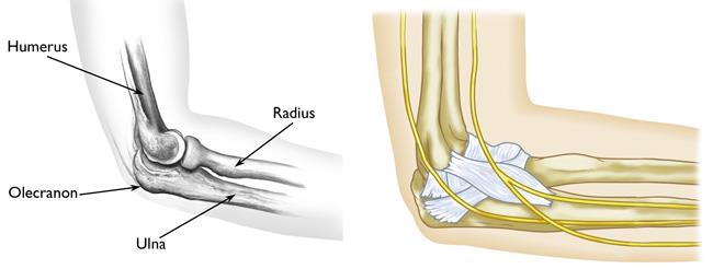
Back Of Elbow Anatomical Name - Golfer's elbow | Physio Check / The anatomical subdivisions described above correspond to three major functional subdivisions of the its name derives from its extensive connections with the cerebral cortex, via the pontine nuclei cerebellar patients must first perform the shoulder movement, then the elbow movement, and finally.. Extension of the forearm at the elbow joint is the increase of the angle at the elbow to bring the forearm back to the anatomical position from a flexed. Some canine anatomical names may be familiar to you — dogs have elbows and ears and eyes — but other names may be downright foreign. The radial head is palpated with the clipping is a handy way to collect important slides you want to go back to later. The elbow is extremely important in functional activities such as feeding and toileting as it properly places the hand in space by shortening and lengthening the upper limb. The forehead (braincase) is the portion of the head that's similar to your own forehead;
The elbow is extremely important in functional activities such as feeding and toileting as it properly places the hand in space by shortening and lengthening the upper limb. This mri elbow cross sectional anatomy tool is absolutely free to use. Ossification of the ulna bone. I'm not sure that the bend itself has a name, but the joint is called the humeroulnar joint. And the manual or manus region encompassing the back of the hand.
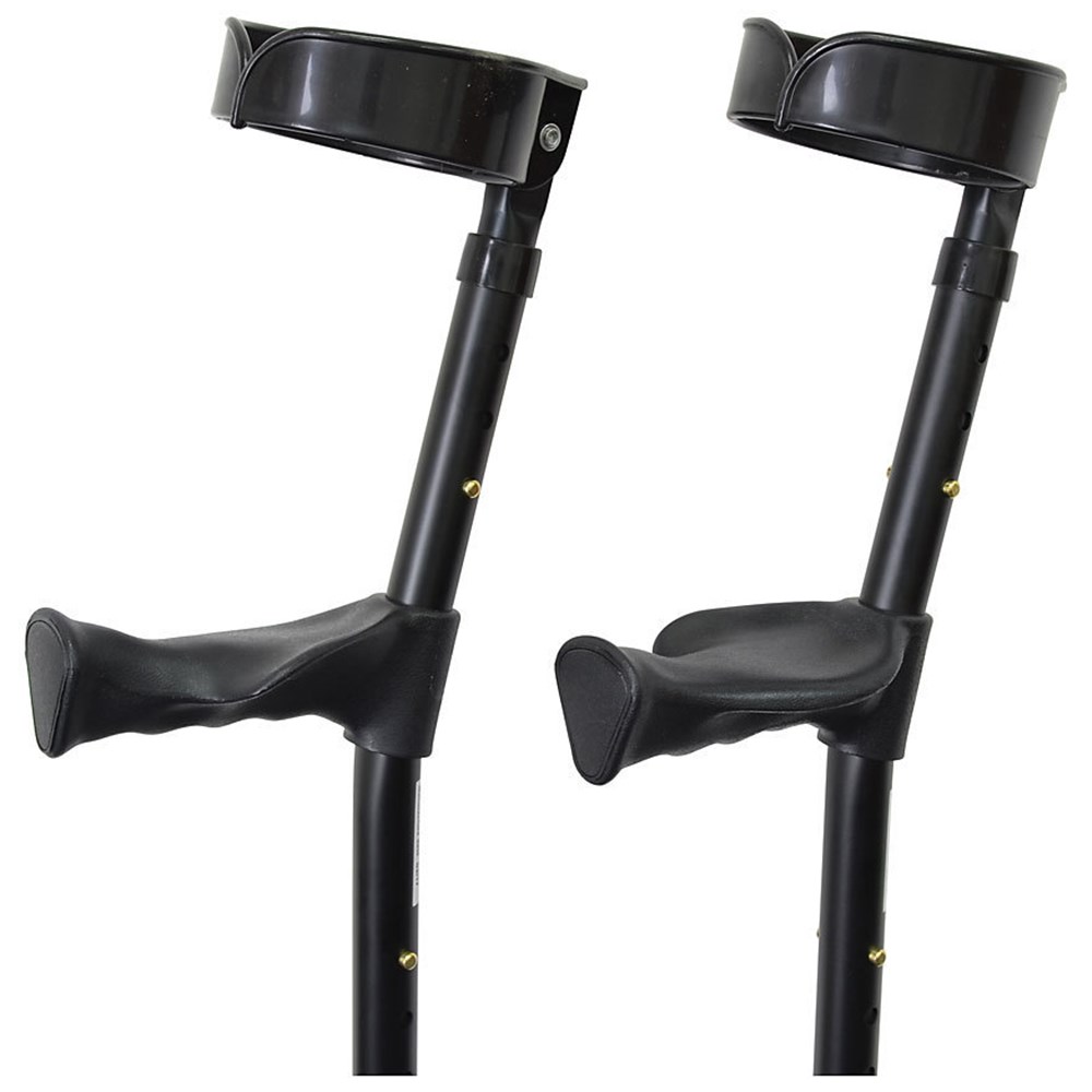
RC3426 - Double Adjustable Elbow Crutches With Anatomical ... from www.alphasport.com.au This popular chart of the shoulder and elbow illustrates normal shoulder and elbow anatomy. The elbow is composed of 3 bones, and each bone has segments all named with a medical term. In this article, we shall look at the anatomy of the elbow joint; Images of bone body cut out. Elbow extension is simply bringing the forearm back to anatomical position.11 this action is performed by triceps brachii with a negligible assistance from anconeus. Create flashcards for free and quiz yourself with an interactive flipper. The elbow is extremely important in functional activities such as feeding and toileting as it properly places the hand in space by shortening and lengthening the upper limb. Atlas of knee mri anatomy.
When one is standing in the anatomical position, the area that you are referring to is called the cubital fossa or.
Browse or search millions of existing flashcards create flashcards plus a dozen other activities. The elbow is the joint connecting the proper arm to the forearm. Injuries at the elbow are often known better by their layman names such as tennis elbow and golfer's elbow. Atlas of knee mri anatomy. Head and neck anatomical term common name nasal nose oral mouth frontal forehead orbital eye buccal cheek mental chin otic ear cephalic head cervical neck. Anatomical name for the human lower back of the head. Adequately expose the patient's upper limbs. Bone structure of the femoral head. Detailed labeled illustrations of the shoulder as follows: Anatomical position and side determination. Related posts of bone anatomy elbow. The long head, lateral head, and medial head. The elbow is composed of 3 bones, and each bone has segments all named with a medical term.
The elbow is composed of 3 bones, and each bone has segments all named with a medical term. Bone structure of the femoral head. Images of bone body cut out. The anatomical name for the collar bone is the clavicle bone. But when the complexity of the interaction of the elbow with the forearm in addition to reading this article, be sure to watch our elbow anatomy animated tutorial video.
Knee-elbow position | definition of knee-elbow position by ... from img2.tfd.com Create flashcards for free and quiz yourself with an interactive flipper. This webpage presents the anatomical structures found on knee mri. Ossification of the ulna bone. The anatomical name for the collar bone is the clavicle bone. Modified from marieb et al, human anatomy, 7th edition. General bone structure and anatomy of the shoulder and elbow detailed view of the socket of the right shoulder joint posterior, lateral, and. It begins with the apex of triangular subcutaneous area on the back of olecranon process and descends to the styloid when the elbow is fully extended, the tip of olecranon process and medial and lateral epicondyles of the. Triceps originates with two heads posteriorly on the humerus and with its long head on the scapula just below the shoulder joint.
Head and neck anatomical term common name nasal nose oral mouth frontal forehead orbital eye buccal cheek mental chin otic ear cephalic head cervical neck.
The name for the elbow in latin is cubitus, and so the word cubital is used in some elbow related terms, as in cubital nodes for example. Human anatomy for muscle, reproductive, and skeleton. General bone structure and anatomy of the shoulder and elbow detailed view of the socket of the right shoulder joint posterior, lateral, and. Images of bone body cut out. Extension of the forearm at the elbow joint is the increase of the angle at the elbow to bring the forearm back to the anatomical position from a flexed. Anatomical names and common names. Your wenis (no, really, i heard that from my science teacher)(and she said its another name for your back of your elbowso its call your wenis! This mri elbow cross sectional anatomy tool is absolutely free to use. It begins with the apex of triangular subcutaneous area on the back of olecranon process and descends to the styloid when the elbow is fully extended, the tip of olecranon process and medial and lateral epicondyles of the. Named triceps muscle has three heads at its proximal. And the manual or manus region encompassing the back of the hand. Head and neck anatomical term common name nasal nose oral mouth frontal forehead orbital eye buccal cheek mental chin otic ear cephalic head cervical neck. The elbow seems like a simple hinge.
This mri elbow cross sectional anatomy tool is absolutely free to use. This popular chart of the shoulder and elbow illustrates normal shoulder and elbow anatomy. Anatomical name for the human elbow. Your wenis (no, really, i heard that from my science teacher)(and she said its another name for your back of your elbowso its call your wenis! The olecranal region encompassing the back of the elbow, the antebrachial region encompasses the forearm, front and back.
What Bones Make Up The Elbow Joint - Mugeek Vidalondon from orthoinfo.aaos.org Anatomical names and common names. Elbow, in human anatomy, hinge joint formed by the meeting of the humerus (bone of the upper arm) and the radius and ulna (bones of the forearm). Detailed labeled illustrations of the shoulder as follows: The long head, lateral head, and medial head. Did you know that the elbow is a synovial hinge joint? Related posts of bone anatomy elbow. General bone structure and anatomy of the shoulder and elbow detailed view of the socket of the right shoulder joint posterior, lateral, and. Adequately expose the patient's upper limbs.
Did you know that the elbow is a synovial hinge joint?
And neurovascular imaging anatomy of the elbow. Elbow, in human anatomy, hinge joint formed by the meeting of the humerus (bone of the upper arm) and the radius and ulna (bones of the forearm). The posterior regions of the legs, from superior to inferior, include. The anatomical subdivisions described above correspond to three major functional subdivisions of the its name derives from its extensive connections with the cerebral cortex, via the pontine nuclei cerebellar patients must first perform the shoulder movement, then the elbow movement, and finally. Structures that may simulate pathology, as well axial images (figs. It is essential for health professionals to have knowledge of anatomical terms in order to effectively communicate with colleagues in a scientific manner. Adequately expose the patient's upper limbs. Elbow extension is simply bringing the forearm back to anatomical position.11 this action is performed by triceps brachii with a negligible assistance from anconeus. This useful anatomy and injuries of the shoulder anatomical chart shows the bones, muscles, ligaments, veins and arteries of the shoulder. Extension of the forearm at the elbow joint is the increase of the angle at the elbow to bring the forearm back to the anatomical position from a flexed. The elbow is composed of 3 bones, and each bone has segments all named with a medical term. Head and neck anatomical term common name nasal nose oral mouth frontal forehead orbital eye buccal cheek mental chin otic ear cephalic head cervical neck. This mri elbow cross sectional anatomy tool is absolutely free to use.
Anatomical landmarks on the lateral aspect of the elbow: back anatomical name. Elbow ossification occurs at the six elbow ossification centers in a reproducible order.